Ocular Image Analysis Research Group
School of Advanced Medical Technologies in Medicine (ATiM)
About
The first session was held up in autumn 2009 and has been going on till now on Sundays at 2.00 pm.
ACHIVEMENT
Ocular Image Analysis Research Group
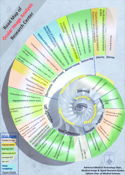
Goals
- Modeling
- Preprocessing
- Segmentation
- feature extraction
- and classification.

ACHIVEMENT
- 3rd rank in the OPTIMA Retinal Cyst Segmentation Challenge, MICCAI 2015 to extract the cysts from OCT images by Dr. Esmaeeli.
- The Winner of ICASSP 2011 Travel Grant
- The Winner of IEEE Signal Processing Society (SPS) Grant in Proc. 2010 IEEE International Conference on Image Processing (ICIP)
- The Winner of IEEE ICIP Student Author Participation Award in Proc. 2009 IEEE International Conference on Image Processing (ICIP)
- The winner of research Scholarship of TUBITAK 2216, 2014.
- Election as the best booth in the second annual conference of AMT faculty.

Open positions
- portable ocular imaging device
- Mathematical modeling of 3-D OCT images
- Geometrical modeling of 3-D OCT images
- Multivariate statistical modeling of 3-D OCT images
- Application of MCA for ocular Images
- Classification of ocular images using the Optimum basic functions
- Classification of vein and artery based on pulsation in angiographic images
- Segmentation of retinal layers of OCT images in patients with retinal holes
- Combination the information of OCT and ERG
- High Order Spectral (HOS) analysis of OCT images to analyze the layers
- Comparison of OCT in left and right eyes in diabetic patients
- Glaucoma diagnosis based on RNFL tissue
- Comparison of GCL and RNFL thickness based on OCT
- Detection occlusion of vessel based on OCT
- Comparison of confocal microscopy with OCT projection results in corneal imaging
- Vessel segmentation in EDI OCT
- Classification of retinal vessels by point elongation method
- Automatic detection foci in OCT image
- Finding the optimal basis of retinal layers in OCT images
- Micro aneurysm detection in OCT images

Articles
PUBLICATION

The Ocular Images Analysis Research Group provides an environment to gather researchers which are interested in this field. The main goal of this research group is to present efficient algorithms for automatic and semi-automatic ocular image analysis. The first session was held up in autumn 2009 and has been going on till now on Sundays at 2.00 pm.
Read MoreTeam
Dr. Hossein Rabbani
manager
Associate professor at Isfahan University of Medicine Science IUMS, senior member of IEEE, founder of Medical Image & Signal Processing(MISP)
Dr. Raheleh Kafieh
Associate professor at IUMS
Title of PhD. thesis Combination of graph based and space-frequency methods in analysis of optical coherence tomography (OCT) images
Dr. Zahra Amini
Associate professor at IUMS
Title of PhD. thesis A new model based on multivariate gaussization of OCT
Mohammadreza akhlaghi
Associate Professor of Retina & Vitreous
School of Medicine Isfahan Eye Research Center Isfahan University of Medical Sciences
Contact
- Iran
- Eight corner Cottage (former library), next to the EDC building, Isfahan University of Medical Sciences


